
Optical tweezers
The optical tweezers experiment by Mills et al. (2004) was simulated by J. Siguenza (PhD Student) by using the YALES2BIO platform. The configuration is symmetric and only half of the cell is shown in the movie. A force is applied to two opposite circular region over the red blood cell initially at rest in order to mimic the silica sphere used in the experiment. After the transient is evacuated, a stationary deformation is obtained which can be compared to the experimental data. Using the Skalak et al. (1973) strain energy function allows a very good representation of the length and radius of the stretched red blood cell.
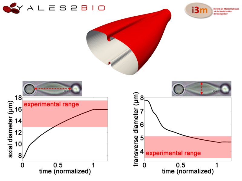
MOVIE: Model of red blood cells stretched by optical tweezers (the RBC is cut in half to allow better observation of the shape)
More details can be found in Siguenza, Mendez and Nicoud, BMM, 16(5), 2017. See also Siguenza et al., JCP, 322, 2016 for more details about the numerical method and benchmark test cases.